Published on: February 6, 2024
Author: Admin
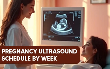
Pregnancy ultrasounds, also known as sonograms are an important aspect throughout the pregnancy period. Using sound waves, this diagnostic approach is used to develop the images of the foetus in the uterus. They are generally performed to detect the health of the unborn baby and the mother’s reproductive organs.
Pregnancy Ultrasound Schedule

Listed below are the most common pregnancy ultrasounds week by week:
- An early pregnancy scan or dating scan (8 – 11 weeks)
- A nuchal translucency (NT) scan (11 – 14 weeks)
- An early anomaly scan (14 – 18 weeks)
- A fetal anomaly scan (19 – 23 weeks)
- A fetal well-being and growth scan (24 – 42 weeks)
Early Pregnancy Scan or Dating Scan (8 – 11 weeks)
An early pregnancy scan is performed to confirm the conception of the child. During this scan, the expected date of delivery (EDD) is determined. Also, the fetal heart rate is detected.
In addition, the blood test known as a non-invasive prenatal test (NIPT) is recommended to determine the possibilities of Down syndrome.
Need expert advice? Consult our doctors now!
Call Now: 80047 80048Nuchal Translucency Scan (NT) (11 – 14 weeks)
A nuchal translucency scan or NT scan is a non-invasive ultrasound to screen the chances of genetic medical conditions and Down syndrome during pregnancy. Generally, this scan is performed abdominally. In some cases, an internal scan is suggested.
The NT scan is performed to:
- Confirm the developing infant’s heart rate.
- Determine the pregnancy duration more accurately.
Note: The NT scan may not be significant for pregnancies conceived by IVF, since the dates are calculated from the embryo transfer.
- Measure the length of the developing infant.
- Examine the number of existing gestations in the pregnancy and analyse whether they are sharing the placenta, which requires close monitoring throughout the pregnancy.
- Analyse the structure of the developing infant, including the level of amniotic fluids.
- Determine the possibilities of chromosomal medical conditions such as:
- Down syndrome (trisomy 21)
- Trisomy 18 (Edwards syndrome)
- Trisomy 13 (Patau syndrome)
Note: If any abnormalities are found in the developing infant, the healthcare provider will provide a follow-up discussion and counselling for further options.
Early Anomaly Scan (14 – 18 weeks)
An early anomaly sonogram pregnancy schedule is performed to determine the significant physical abnormalities in the unborn child.
During this scan, the developing baby’s physical structure is examined, including:
- Limbs and bones
- Internal organs such as the brain, heart, kidneys, and spinal cord
The expectant mother's uterus is also analysed for any chances of uterine medical conditions.
Fetal anomaly scan (19 – 23 weeks)
A fetal anomaly scan is a systematic sonogram that examines the developing baby from head to toe and determines whether the structures are appropriate as per the gestational age. Most abnormalities are well distinguished in this scan, if any.
Note: If there are any abnormalities found in the developing infant, a follow-up assessment or sonogram is suggested to confirm the condition. If it is established, discussion and counselling are provided by the healthcare provider for further options.
During this ultrasound:
- The blood flow from the placenta and the position of the placenta is examined.
- The length of the cervix is measured to analyse the risk of preterm labour.
Fetal Well-Being and Growth Scan (24 – 42 weeks)
A fetal well-being or growth scan is the significant scan after 24 weeks of pregnancy. During this scan, the well-being of the developing baby is closely monitored.
The following measurements and examinations are done during this scan:
- The baby’s head, thigh, and abdomen are measured to analyse the appropriate weight as per the gestational age.
- The level of amniotic fluid around the developing baby is measured.
- The baby’s blood vessels and the blood flow through the umbilical cord are examined through the Doppler scans.
Note: During this period, the placement of the baby in the mother’s womb plays a vital role in planning the delivery.
Types of Ultrasounds during Pregnancy
The most advanced ultrasound techniques are used if the physician requires more detailed images of the developing baby. These are helpful for the physicians to make a clear diagnosis if there are any abnormalities found in the developing infant.
Transvaginal Ultrasound:
A transvaginal ultrasound is helpful to develop a clearer image of the developing baby. This ultrasound is typically used in the first trimester of pregnancy. To perform the test, a small scan probe is inserted into the vagina to capture the images.
3-D Ultrasound:
A 3-D ultrasound involves the ultrasound schedule pregnancy to assist the physician in analysing the height, depth, and width of the developing baby and the expectant mother’s reproductive organs. This sonogram plays a key role in establishing abnormalities during pregnancy.
A traditional 2-D and 3-D ultrasound are similar in performing the procedure. However, they differ in using software and a probe to develop the 3-D images. In addition, 3-D ultrasounds can be handled only by trained professionals.
4-D Ultrasound:
A 4-D ultrasound is also known as a dynamic 3-D ultrasound. Unlike 2-D and 3-D sonograms, 4-D sonograms create a moving video of the developing baby in the mother’s uterus. This is helpful to develop a clear image of the baby’s structures and movements. A 4-D ultrasound is performed using special equipment, unlike other ultrasounds.
Fetal Echocardiography:
A fetal echocardiography is suggested by the physicians if they suspect the developing baby in the uterus has a congenital heart condition. The examination usually consumes more time to complete.
The test is useful to capture in-depth images of the developing baby’s heart, including its shape, size, and structure. It helps the physician closely monitor the functionality of the unborn baby’s heart and diagnose the condition more specifically.
Doppler Ultrasound:
A Doppler ultrasound is performed to determine the blood flow of the baby through its blood vessels. Generally, most Doppler ultrasounds are performed in the third trimester of pregnancy.
Throughout the pregnancy period, it is always recommended not to miss the pregnancy scan dates. Following routine checkups will help to monitor the health of both the mother and the baby.
Purpose of Ultrasounds during Pregnancy
The pregnancy ultrasound schedule is generally done at three trimesters namely first, second, and third. Pregnant women have ultrasounds throughout the conception period for numerous reasons. They are:
First Trimester (0 – 12 Weeks)
- Confirm the conception of the child.
- Analyse the fetal heart rate.
- Assess the gestational age of the developing infant.
- Analyse the expected date of delivery.
- Examine the number of foetuses present in the womb.
- Analyse the health of reproductive organs such as the cervix, ovaries, placenta, and uterus.
- Detect the ectopic pregnancy or abortion.
- Determine any abnormalities in the developing infant.
Second Trimester (12 – 24 Weeks)
- Monitor the unborn baby’s growth and position (transverse, breech, optimal, or cephalic).
- Confirm the number of fetuses present in the womb.
- Determine if there are any Down syndrome in the developing infant.
- Examine the health of the placenta to analyse if there are any issues related to placenta previa and placental abruption.
- Examine the possibilities of Down syndrome.
- Determine the possibilities of birth defects or congenital abnormalities.
- Analyse the possibilities of structural abnormalities in the developing infant.
- Examine the blood flow in the umbilical cord.
- Monitor the amount of amniotic fluid.
- Determine if the developing infant in the womb gets enough oxygen.
- Analyse the cervix length.
- Analyse an intrauterine death.
Third Trimester (24 – 40 Weeks or Childbirth)
- Determine the fetal anatomy in detail.
- Examine the fetal and placental position.
- Measure the fetal weight and size including head, femur, and abdomen.
- Monitor the fetal movements and breathing levels.
- Measure the amount of amniotic fluid.
- Analyse the blood flow from the umbilical cord.
Reasons for More Ultrasounds during Pregnancy
There are several reasons for your physician to suggest more ultrasounds during pregnancy. Some of the reasons include:
- A medical condition related to the expectant mother’s reproductive organs.
- The developing baby is smaller in size for its gestational age.
- Suspecting intrauterine growth restriction (IUGR).
- Abnormalities with the placenta such as placental abruption or placenta previa.
- Multiple gestations.
- The developing baby is in a breech position.
- Excessive level of amniotic fluid around the baby. The condition is called polyhydramnios.
- Insufficient level of amniotic fluid around the baby. The condition is called oligohydramnios.
- The expectant mother experiences preeclampsia or gestational diabetes.
- The developing baby in the mother’s uterus has a congenital disorder.
Ultrasounds or sonography of pregnancy week by week are typically safe for both the developing baby and the expectant mother.
Many medical associations suggest that healthcare professionals perform ultrasounds throughout the pregnancy when they are medically required.
Repeated scans are not required when the pregnancy is uncomplicated and at lower risk.